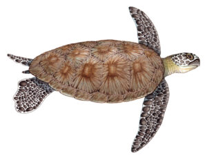
Green sea turtle Chelonia mydas
Green sea turtle Chelonia mydas completed for UKOTCF Montserrat
Yellow line arrow crab Stenorhyncus seticornis
Yellow line arrow crab Stenorhyncus seticornis completed for UKOTCF Montserrat
Carribean hermit crab Coenobita clypeatus
Carribean hermit crab Coenobita clypeatus completed for UKOTCF Montserrat
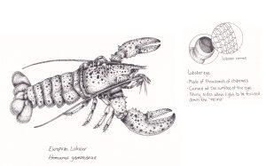
European lobster Homarus gammarus with eye detail
European lobster Homarus gammarus with eye detail showing curved lens Illustration from “30 Animals that made us smarter” by Patrick Ayree (BBC Books)
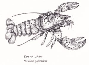
European lobster Homarus gammarus
European lobster Homarus gammarus Illustration from “30 Animals that made us smarter” by Patrick Ayree (BBC Books)
Blue mussels Mytilus edulis
Blue mussels Mytilus edulis with byssal threads on wooden piling. Illustration includes barnacles and limpets, and some bladder wrack seaweed. Illustration from “30 Animals that made us smarter” by Patrick Ayree (BBC Books).
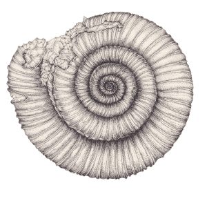
Ammonite Dactylioceras commune
Ammonite Dactylioceras commune fossil similar to those collected by David Attenborough as a child
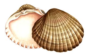
common cockle Cerastoderma edule shell
common cockle Cerastoderma edule pair of shells from one animal, cut to white
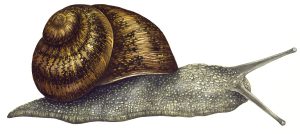
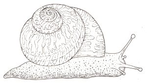
Garden snail Helix aspera
Garden snail Helix aspera with grey body and yellow and brown shell cut to white
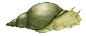
Giant pond snail Lymnaea stagnalis
Giant pond snail Lymnaea stagnalis cut to white
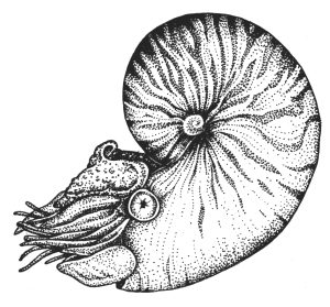
Nautilus pompilius
Nautilus pompilius pen and ink stippled illustration
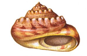
Topshell Gibbula umbilicalis
Topshell sketch in colour cut to whtie
Garden snail Helix aspera 2
Garden snail Helix aspera line drawing in pen and ink cut to white
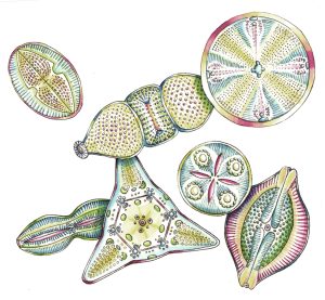
Diatoms
Diatom group showing diverse shapes and colours of these organisms
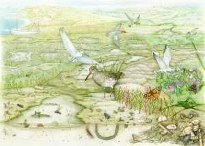
Saltmarsh landscape
Salt marsh landscape showing mud and a cross section of the snails and invertebrates who live here, wading and predatory birds above in the glasswort and aquatic plants; further away from the salt estuary are soft cliffs and the distant sea
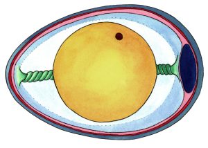
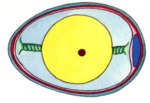
Schematic Diagram of Duck Egg
Stylized diagram of the duck egg showing chalaza, yolk, white, germination, membranes, and air pocket
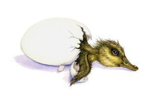
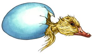
Mallard duckling just hatched Anas platyrhynchos
Mallard Anas platyrhynchos duckling in the process of hatching from its egg, half in half our and exhausted with wet feathers
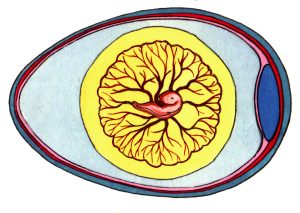
Diagram of Duck Egg development Day 5
Stylized diagram of the development of a duck egg from being laid until after hatching day five embryo separates from the yolk, blood vessel network grows and branchesm eye starts to get a colour.
Diagram of Duck Egg development Day 8
Stylized diagram of the development of a duck egg from being laid until after hatching day eight blood vessels carry waste and oxygen, eyelids start to cover eye, beak and egg tooth more clear.
Diagram of Duck Egg development Duckling chick hatching out still half in its shell
Stylized diagram of the development of a duck egg from being laid until after hatching duckling hatching half in half out of shell with prominent egg tooth
Diagram of Duck Egg development Hatched duckling chick
Stylized diagram of the development of a duck egg from being laid until after hatching hatched duckling chick
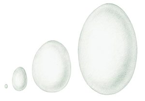
Comparison of egg sizes
Pencil sketch comparing size of humming bird, ostrich, hen, and elephant bird
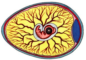
Diagram of Duck Egg development Day 1
Stylized diagram of the development of a duck egg from being laid until after hatching Day one yolk held in place with chalaza, germinal disc where fertilized chick starts to grow
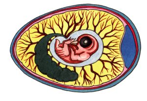
Diagram of Duck Egg development Day 12
Stylized diagram of the development of a duck egg from being laid until after hatching day twelve feathers start to show eye covered with lid claws formed