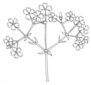
Dichasial cyme
Dichasial cyme diagram showing the pattern of this flowering form
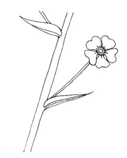
Disc floret
Disc floret diagram with seed attached and stamens and pistil on show
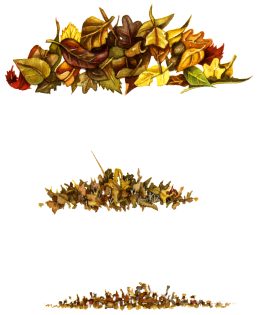
Dead leaf decomposition
Dead leaves rotting and decomposing into compost from newly fallen to decomposed and down to small leaf patricles like compost
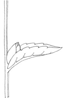
Diagram of a bract
Diagram of a bract with little flower growing from behind the bract
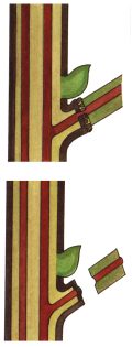
Bulbil diagram
Bulbil diagram showing how it emerges at the junction of the stem and leaf
Auxin and growth diagram
Auxin diagram showing how applying auxin to the top of a plant encourages vertical rather than lateral growth
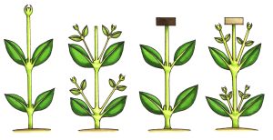
Abscission diagram
Abscission diagram showing ho w the abscission layer lets the leaf fall off without exposing the plant to pathogens
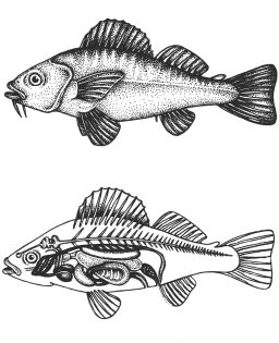
Fish anatomy diagram
Fish anatomy diagram simplified exterior and interior cross section view
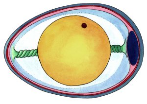
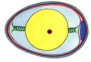
Schematic Diagram of Duck Egg
Stylized diagram of the duck egg showing chalaza, yolk, white, germination, membranes, and air pocket
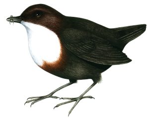
Dipper Cinclus cinclus
Dipper eating aquatic invertebrate
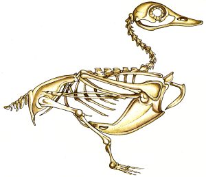
Diagram of a duck skeleton
Stylized diagram of the skeelton of a duck
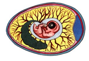
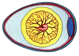
Diagram of Duck Egg development Day 1
Stylized diagram of the development of a duck egg from being laid until after hatching Day one yolk held in place with chalaza, germinal disc where fertilized chick starts to grow
Diagram of Duck Egg development Day 12
Stylized diagram of the development of a duck egg from being laid until after hatching day twelve feathers start to show eye covered with lid claws formed
Diagram of Duck Egg development Day 27
Stylized diagram of the development of a duck egg from being laid until after hatching Day 27 hatchling squashed inside egg ready to hatch, air cell still shows
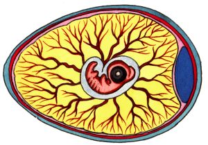
Diagram of Duck Egg development Day 5
Stylized diagram of the development of a duck egg from being laid until after hatching day five embryo separates from the yolk, blood vessel network grows and branchesm eye starts to get a colour.
Diagram of Duck Egg development Day 8
Stylized diagram of the development of a duck egg from being laid until after hatching day eight blood vessels carry waste and oxygen, eyelids start to cover eye, beak and egg tooth more clear.
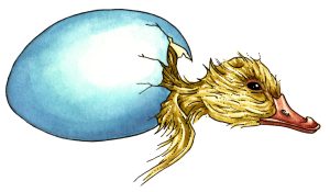
Diagram of Duck Egg development Duckling chick hatching out still half in its shell
Stylized diagram of the development of a duck egg from being laid until after hatching duckling hatching half in half out of shell with prominent egg tooth
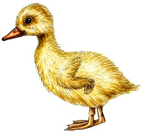
Diagram of Duck Egg development Hatched duckling chick
Stylized diagram of the development of a duck egg from being laid until after hatching hatched duckling chick
Diagram of the cross section of a bird bone
Bird bone cross section showing cross bracing for strength and hollow structure
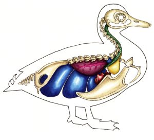
Diagram of the internal anatomy of a duck
Stylized diagram of the internal organs and anatomy of a duck within a pen outline